Day 2 :
Keynote Forum
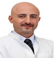
Sebouh Kassis
Al Zahra Hospital, UAE
Keynote: Managing non functioning pituitary neuroendocrine tumors based on the best available evidence
Time : 09:00-09:50
Biography:
Sebouh Kassis is a Neurosurgeon and Spine Surgeon. He has completed his Neurosurgical training from the University Hospital of Liege in Belgium where he was graduated with the Highest Distinction Award and became holder of Belgian Board Certificate in Neurosurgery. He has also completed a Fellowship program of 3 years in the Neurological Hospital of Lyon, France and is a holder of French Specialty Certificate for both Neurosurgery and Neurology. He has special interest and expertise in the treatment of pituitary and skull base pathologies with fully endoscopic approaches, microsurgical and minimally invasive treatment of diverse spinal pathologies, spinal fusion including deformity correction. He has also committed to the integration of spinal navigation and neurophysiology monitoring in the management of complex spine cases. He is the Head of the Neurosurgery Department of Al Zahra Private Hospital, Dubai.
Abstract:
Non-Functioning Pituitary Neuroendocrine Tumors (NF-PitNETs) represent about half of all pituitary neuroendocrine tumors. Being clinically silent, NF-PitNETs are usually discovered as macroadenomas compressing the optic chiasma, the pituitary gland or/and the pituitary stalk. Surgical removal remains the treatment of choice considering almost the absence of an effective medical treatment in contrast to most functioning pituitary tumors. Although the vast majority of NF-PitNETs are benign, their treatment remains a challenge considering that about 50% present with cavernous sinus invasion at the time of diagnosis which limits the radical removal. It is estimated that about half of the patients with invasive NF-PitNETs present regrowth of the residual tumor and about 15% present growth after gross total removal without residue. Inspired from the recent 2017 WHO classification and numerous studies, the term (high risk pituitary adenoma) evolved in the recent years; this includes tumors with increased cell proliferation and signs of invasive growth evaluated by MRI and/ or histology. Subsequently, the optimal treatment strategy of NF-PitNETs nowadays should not only consider the choice of surgical technique but also identifying factors which help predicting the risk of recurrence. In this presentation, the author exposes the current surgical techniques and discusses the predictive factors of recurrence and the different management modalities in such cases.
Keynote Forum
Gajendra Prasad Rauniar
B.P.Koirala Institute of Health Sciences, Nepal
Keynote: Therapeutic drug monitoring of antiepileptic drugs in BPKIHS, Dharan
Time : 09:50-10:40

Biography:
Gajendra Prasad Rauniar is currently working as a Professor in the Department of Clinical Pharmacology and Therapeutics at B.P. Koirala Institute of Health Sciences, Dharan, Nepal. He has published more than 60 scientific papers in national and international journals. He is currently doing research in neuropharmacology related clinical trials as well as drug utilization.
Abstract:
Keynote Forum
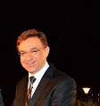
Hischam Bassiouni
Klinikum St. Marien Amberg, Germany
Keynote: Lesion-tailored approaches in spinal surgery
Time : 11:00-11:50
Biography:
Hischam Bassiouni is an Associate Professor of Neurosurgery. He is the Director of two Neurosurgical Clinics at two major academic teaching hospitals, (Klinikum Amberg and Klinikum Weiden) in Bavaria, Germany. He is also the Member of German Neurosurgical Society, European Neurosurgical Society and German Skull Base Society. He had his Neurosurgical training at University Hospital Aachen and University Hospital Essen, Germany. His neurosurgical and scientific sub-specializations include neuro-oncology, neurovascular surgery, skull base surgery and neuro-pediatric surgery. He is the first author of 13 publications in highranked Neurosurgical journals and has authored several chapters in international neurosurgical reference books.
Abstract:
- Spine and Spinal Disorders|Spine Surgery | Neuropharmacology
Location: Dubai
Chair
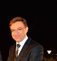
Hischam Bassiouni
Klinikum St. Marien Amberg, Germany
Co-Chair
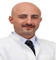
Sebouh Kassis
Al Zahra Hospital, UAE
Session Introduction
Basem Awad
Mansoura University Hospital, Egypt
Title: Lateral extracavitary approach to spinal tumors
Time : 11:50-12:20
Biography:
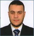
Basem I. Awad is an Assistant Professor of Neurosurgery from Egypt, currently working at Mansoura University School of Medicine, in Mansoura, Egypt. He is the Educational Neuro officer at the AOSpine Egypt Council Board. He has completed his Master Degree of Surgery at Mansoura University, Egypt and Doctorate Degree at Joint between Case Western Reserve University, Cleveland, OH and Mansoura University. He also received the Crockard International Spine Fellowship at Cleveland Clinic and the AOSpine International Fellowship at the Center for Spinal Disorders, CO, USA. Recently, he completed Bioinformatics PostDoc Fellowship at Luxembourg Center for Systems Biomedicine, University of Luxembourg. Dr. Awad is also member of many international socities e.g. American Association of Neurosurgery (AANS), Congress of Neurosurgery (CNS), AOSpine, and North Americam Spine Society (NASS). He was selected to be on of the EDITORIAL BOARD for the Global Spine journal and World NEUROSURGERY Journal. His neurosurgical and scientific subspecializations includes spinal disorders and surgery, spinal trauma, spinal cord injury, neuro-oncology.
Abstract:
The surgical management of pathology involving the ventral aspect of the thoracic and upper lumbar spine is typically challenging. Thoracotomy provides direct ventral exposure of the spine and spinalcord. However the approach related morbidities could be markedly significant while a separate dorsal approach may be required for instrumentation. The Lateral Extracavitary Approach (LECA) is a dorsolateral approach that provides lateral and ventral access to thoracic and upper lumbar spine without entrance into the pleural cavity. By remaining extra pleural, the LECA avoids the complications noticed previously with thoracotomy. Neural decompression, tumor removal and fixation can all be accomplished via LECA, which makes it an invaluable tool in spinal surgery. This technical advantage has led to excellent neurological outcomes with nearly 75% of patients described in the literature revealing neurological improvement. In the present study, we reviewed 15 patients with spinal tumors treated with anterior and posterior resection and reconstruction from a single posterior approach. Pre- and post-operative neurological condition means blood loss, length of hospital stay after surgery and complications related directly to surgery were analyzed. Pre- and post-operative Computed Tomography (CT) scans and Magnetic Resonance Imaging (MRI) scans were evaluated. Our results showed neurological improvement in 69.2%, 29.2% experienced no change and 1.5% reported worse condition. Mean blood loss was 2,134 mL and hospital stay was 7.2 days. Total complication rate were 15.5%. In conclusion the adequate neural decompression combined with anterior and posterior column reconstruction is feasible through lateral extracavitary approach using a single posterior skin incision. Minimally Invasive (MIS) approaches are now being applied in all areas of the spine surgeries including LECA. MIS LECA approach is purported to have decreased operative time, reduced blood loss, less tissue dissection, less perioperative pain and earlier mobility.
Basem Awad
Mansoura University Hospital, Egypt
Title: Minimally invasive approach for intradural extra-medullary spinal tumors
Time : 12:20-12:50
Biography:
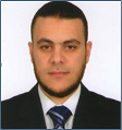
Basem I. Awad is an Assistant Professor of Neurosurgery from Egypt, currently working at Mansoura University School of Medicine, in Mansoura, Egypt. He is the Educational Neuro officer at the AOSpine Egypt Council Board. He has completed his Master Degree of Surgery at Mansoura University, Egypt and Doctorate Degree at Joint between Case Western Reserve University, Cleveland, OH and Mansoura University. He also received the Crockard International Spine Fellowship at Cleveland Clinic and the AOSpine International Fellowship at the Center for Spinal Disorders, CO, USA. Recently, he completed Bioinformatics PostDoc Fellowship at Luxembourg Center for Systems Biomedicine, University of Luxembourg. Dr. Awad is also member of many international socities e.g. American Association of Neurosurgery (AANS), Congress of Neurosurgery (CNS), AOSpine, and North Americam Spine Society (NASS). He was selected to be on of the EDITORIAL BOARD for the Global Spine journal and World NEUROSURGERY Journal. His neurosurgical and scientific sub specializations includes spinal disorders and surgery, spinal trauma, spinal cord injury, neuro-oncology.
Abstract:
Arman Rahimmi
Kurdistan University of Medical Sciences, Iran
Title: Are anti-oxidants & anti-inflammatory compounds good choices for curing Parkinson’s disease?
Time : 14:00-14:30
Biography:
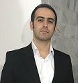
Arman Rahimmi is a PhD student of Molecular Medicine in Kurdistan University Medical Sciences, Sanandaj, Iran. His research works has been focused on molecular nature of neurodegenerative disease especially Parkinson’s disease. His studies include the role of oxidative stress and inflammation in those diseases and evaluate potentials of antioxidants and anti-inflammatory compounds for treating them.
Abstract:
Parkinson’s Disease (PD) is a progressive neurodegenerative disorder, which is considered as one of the most prevalent diseases of Central Nervous System (CNS). Its clinical signs include both motor (resting tremor, rigidity and bradykinesia) and mental disorders (cognitive problems, behavioral impairments and dementia). These clinical symptoms are mainly the consequents of progressive loss of dopaminergic (DAergic) neurons in brain, especially those of Substantia Nigra (SN) and Striatum (ST). Accordingly, current PD therapies focus on maintaining dopamine levels of brain at normal range. However, this approach is fairly useful to control and manage Parkinson’s disease, it has some disadvantages. Firstly, patients need higher doses of drugs over time which it implies some serious side effects such as psychosis, motor fluctuations, and dyskinesias. Additionally, PD patients under this type of treatment develop a series of dopa-resistant motor symptoms (speech impairment, abnormal posture, and gait and balance problems) and dopa-resistant non-motor signs (anosmia, sleep disorders, autonomic dysfunction, mood impairment and pain) after a while. In this regard, previous studies indicate that Levodopa and other dopaminergic medications accelerate neuronal degeneration in some parkinsonian brains via production of free radicals and Reactive Oxygen Species (ROS). This is in addition to the main oxidative and inflammatory processes of PD. Literature strongly confirm the role of oxidative stress and inflammation in development and progression of Parkinson’s disease. So that, during the recent years, interest in administration of neuroprotective factors such as brain repairing antioxidants and anti-inflammatory drugs for management of PD is being popular, increasingly. On the other hand, since PD is a chronic and long-lasting disease, it is important to improve life quality and life expectancy of PD patients by appropriate medications. According to the above literature, it is important to understand the mechanism of action of these neuro-protectant factors and investigate the new and more effective ones. Therefore, the objective of this article is to do a comprehensive review on oxidative and inflammatory mechanisms playing role in pathogenesis of PD. We also highlight the studies concerning antioxidant and anti-inflammation therapies for PD and their molecular mechanisms of action.
Stefan Reguli
University Hospital Ostrava Poruba, Czech Republic
Title: Long non-coding RNA analysis in glioblastoma
Time : 14:30-15:00
Biography:
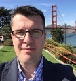
Stefan Reguli is a Neurosurgeon working in the University Hospital Ostrava Poruba, Czech Republic. His main field is Neuro-oncology, especially treating patients with gliomas. He is Coordinator of Ostrava Neuro-Oncology Center and has participated in several studies focused on basic brain tumor research.
Abstract:
Glioblastoma Multiforme (GBM) is the most frequent primary brain tumor of astrocytic origin. The prognosis of GBM patients is very poor with median of overall survival ranging between 12 and 15 months from diagnosis despite conventional therapeutic protocol consisting of maximal surgical resection followed by concomitant chemo-radiotherapy with Temozolomide and adjuvant Temozolomide in monotherapy. Therefore, a lot of financial resources and a great deal of effort are spent in the research of new therapeutic approaches that could prolong the survival of GBM patients. Long non-coding RNAs (lncRNAs) are a relatively new class of noncoding gene regulators playing critical roles in tumor biology, including GBM. From this perspective, lncRNAs seem to be promising therapeutic targets in GBM patients. We performed NGS analysis of fresh-frozen histopathologically confirmed GBM tissues and non-tumor brain tissues obtained from non-dominant anterior temporal cortexes resected during surgery for intractable epilepsy with no signs of dysplastic changes. Informed consent approved by the local Ethical Commission was obtained from each patient before the treatment. rRNA depletion and cDNA library preparation were performed with GeneRead rRNA depletion kit (Qiagen) and NEXTflex rapid directional qRNA-Seq Kit (Bioo Scientific), respectively. Sequencing was done using NextSeq 500 high output kit and NextSeq 500 instrument (both Illumina). Statistical analysis evaluated protein-coding and non-coding RNAs and their sequential variants with non-zero RPKM (reads per kilobase of transcript per million mapped reads) at least in one sample. We used CLC genomic workbench for the alignment and target counts. Targeted regulation of ZFAS1 level has been carried out by the transient transfection of specific siRNA in GBM stable cell lines (A172, U87MG and T98G). The success of transfection and viability were analyzed in vitro using qRT-PCR and MTT assay, respectively. We have demonstrated a dysregulation of many lncRNAs and protein-coding RNAs in GBM tissue in comparison with non-tumor brain tissue. However, the forced down regulation of ZFAS1, one of the most up-regulated lncRNAs in GBM tissue, was not found to have an impact on the viability of GBM cell lines in vitro.
Olaitan Jeremiah
Royal College of Surgeons in Ireland (RCSI), Ireland
Title: Evaluation of the effect of insulin sensitivity- enhancing lifestyle- and dietary- related adjuncts on antidepressant treatment response: A systematic review and meta-analysis
Time : 15:30-16:00
Biography:
Olaitan J. Jeremiah completed her B.Pharm. degree from the Faculty of Pharmacy of Obafemi Awolowo University (OAU), Ile-Ife, Nigeria with a distinction in 2010. She also completed a masters degree in pharmacology from the same university between 2012 and 2014 and was appointed a lecturer in the department of pharmacology, OAU, during the course of her M.Sc programme. Ms. O. J. Jeremiah is presently undertaking her PhD in neuropharmacology at the School of Pharmacy of RCSI, Dublin 2, under the supervision of Dr. Benedict K. Ryan (BSc(Pharm), MPharm, PhD, MPSI).
Abstract:
- Special Session
- Neurology | Neurosurgery |Brain Tumor & Cancer | Neurooncology
Location: Dubai

Chair
Ernesto Miguel Delgado Cidranes
Complutense University Madrid, Spain
Session Introduction
Nibras Al-sumaidaee
Baghdad Neurosurgical Teaching Hospital, Iraq
Title: Glioblastoma multiforme, demographical, clinical features, environmental factors and outcomes after surgical management
Biography:
Nibras Alsumaidaee is a Neurosurgeon. He has completed his training in Neurosurgery from Baghdad Medical Complex (Martyr Gazi Al-hariry for specialized surgical hospital). His interest is mainly towards neuro-oncology, functional neurosurgery.
Abstract:
Abderrahman Omer
Military Hospital-Sudan, Sudan
Title: Neurosurgery in Sudan, current and future challenges
Biography:
Abderrahman Omer is a neurosurgeon at Omdurman Military Hospital, to look after and manage neurosurgical problems as part of the neurosurgical department there, which serves both military and civilian patients. Before starting his career as a neurosurgeon, He joined the ministry of health as a general practitioner and an intern immediately after completing the medical school in the University of Gezira. After having the degree of clinical MD in neurosurgery in 2013,He joined the army hospital and started to work as a neurosurgeon dealing with trauma cases as part of trauma team and nontrauma cases in pediatrics, neuro-oncology and spine cases for simple non complicated cases and later , for the first time in the hospital, to operate on posterior fossa and more complex cases and just few weeks before they did the first transsphenoidal case with ENT team. His ambition is to start more subspecialised surgeries in his hospital and the whole country.
Abstract:
Pankaj R Nepal
B & C Teaching Hospital and Research Center Pvt. Ltd., Nepal
Title: Morphological variation of the confluences of sinuses in head
Biography:
Abstract:
Aim: The aim of the study is to analyze the morphological variation of the confluences of sinuses and propose a classification system.
Method: The study was based on the cross sectional analytical study. Data collection and analysis was done from all the cases of CT Venogram were evaluated in the CT console using the inbuilt software. This Venogram was evaluated using the VRT view of the sinuses and the part of the arterial phage of the angiogram were punched out. Evaluation of the sinuses was done by carefully rotating the venous sinus in all the direction. Evaluation of typical form of the confluence of sinus was identified and variations of the sinuses were evaluated for the rest atypical type.
Result: Total 70 cases were enrolled in the study. Overall the confluence of sinus of Torcula was identified as typical and atypical type. The typical types were further seen as solid or a fenestrated type. The atypical confluences of sinus were seen as (1) Aplastic/hypoplastic transverse sinus, (2) Transverse sinus connecting only with superior sagittal sinus, straight sinus or occipital sinus and (3) Various patterns of occipital sinus either unilateral branching, bilateral branching or no branching. The typical form of the confluences were present in only few cases, however rest were the atypical type. The aplastic or hypoplastic trasverse sinuses were more common in the left side. The presence of occipital sinus in the typical morphology gave the confluence a diamond shaped, however the shape were angled to one side when one of the draining sinus was not connected at Torcula.
Conclusion: Keeping the classification of confluence of sinus in mind could aid in the surgical planning and prevent an advertent injury to the anomalous sinus type.
Ritesh Nawkhare
Bangur Institute of Neurosciences, India
Title: Contiguous involvement of brain and spinal cord by plasma cell granuloma – A rare presentation
Biography:
Abstract:
Plasma cell granuloma of the Central Nervous System (CNS) is a rare entity. Primary intracranial lymphoma was first reported by West et al. in 1980 with around 60 cases reported till date. Previously reported locations include meninges, cerebral hemispheres, ventricles, hypothalamus, sellar region and cerebellum. Intracranial plasma cell granuloma is characterized radiologically by its extra axial, dural based location and homogeneous enhancement with contrast. Owing to it, meningioma, tuberculoma, sarcoidosis and wegners granulomatosis comprise important differential diagnosis. Plasma cell granuloma is generally a benign, non-recurring lesion. Treatment options for plasma cell granuloma include total/subtotal excision, steroids and radiotherapy. Technically inaccessible sites have been treated with biopsy followed by steroid therapy. In majority of cases a single plasma cell granuloma has been reported in the brain or spinal cord. Only two cases of plasma cell granuloma simultaneously involving the brain and spinal cord have been reported. We believe this is the first case showing a single lesion contiguously involving the brain and spinal cord in its extent and was managed surgically and discuss its clinical, radiological and pathological findings along with review of literature.
